EEvlarSoft

DICOM Viewer for Windows

Open the CD from your MRI or CT appointment on a Windows PC, page through every slice, measure what you see and turn the study into a 3D model.

Buy for $29.99
One payment. No subscription.
14-day money-back guarantee.
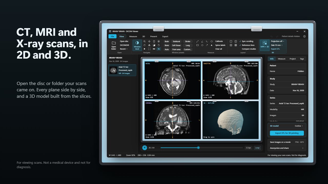
CT, MRI and X-ray scans, every plane side by side with a 3D model.
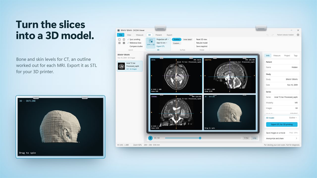
Turn the slices into a 3D model and export it as STL.
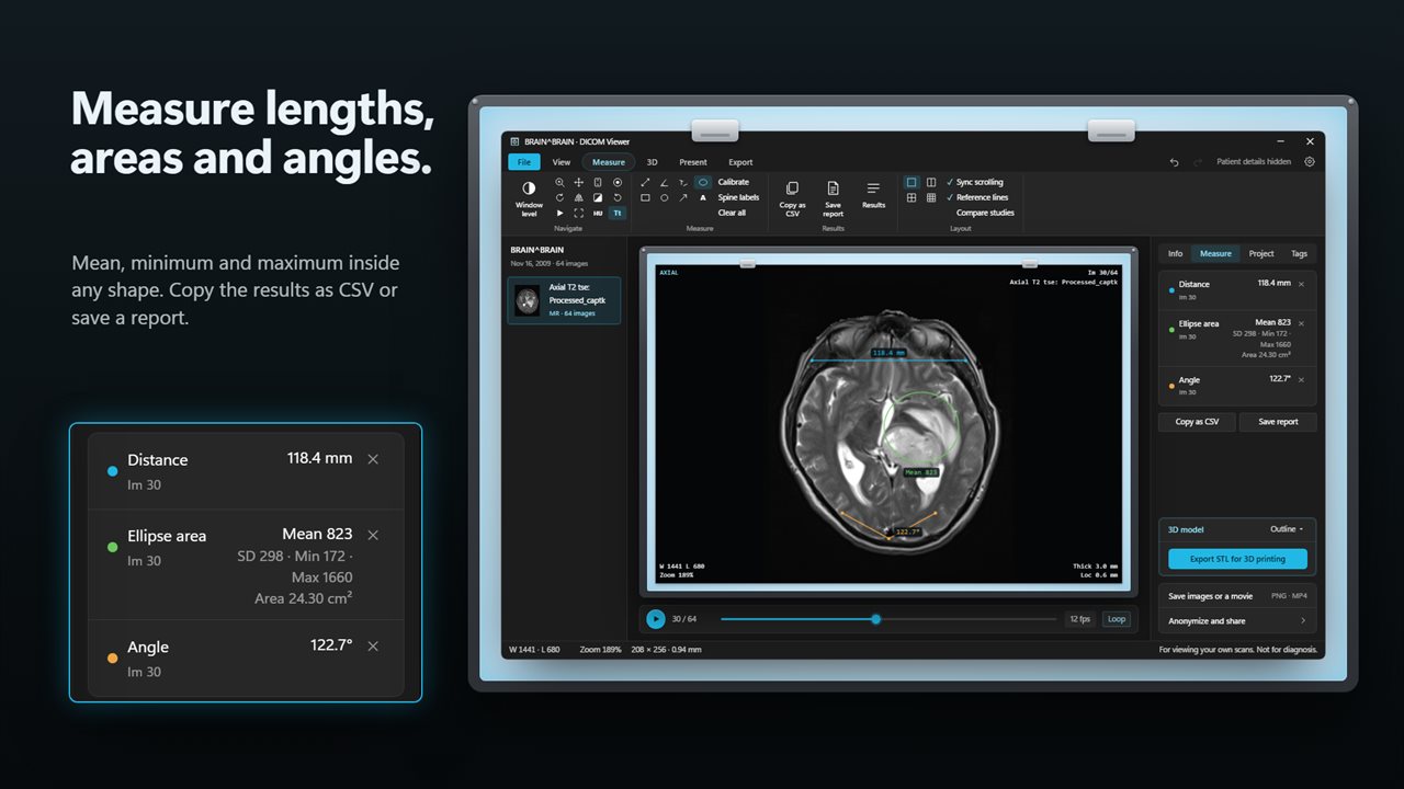
Measure lengths, areas and angles, and copy the results as CSV.
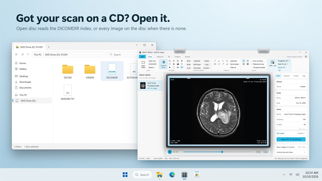
Open the CD your scan came on.
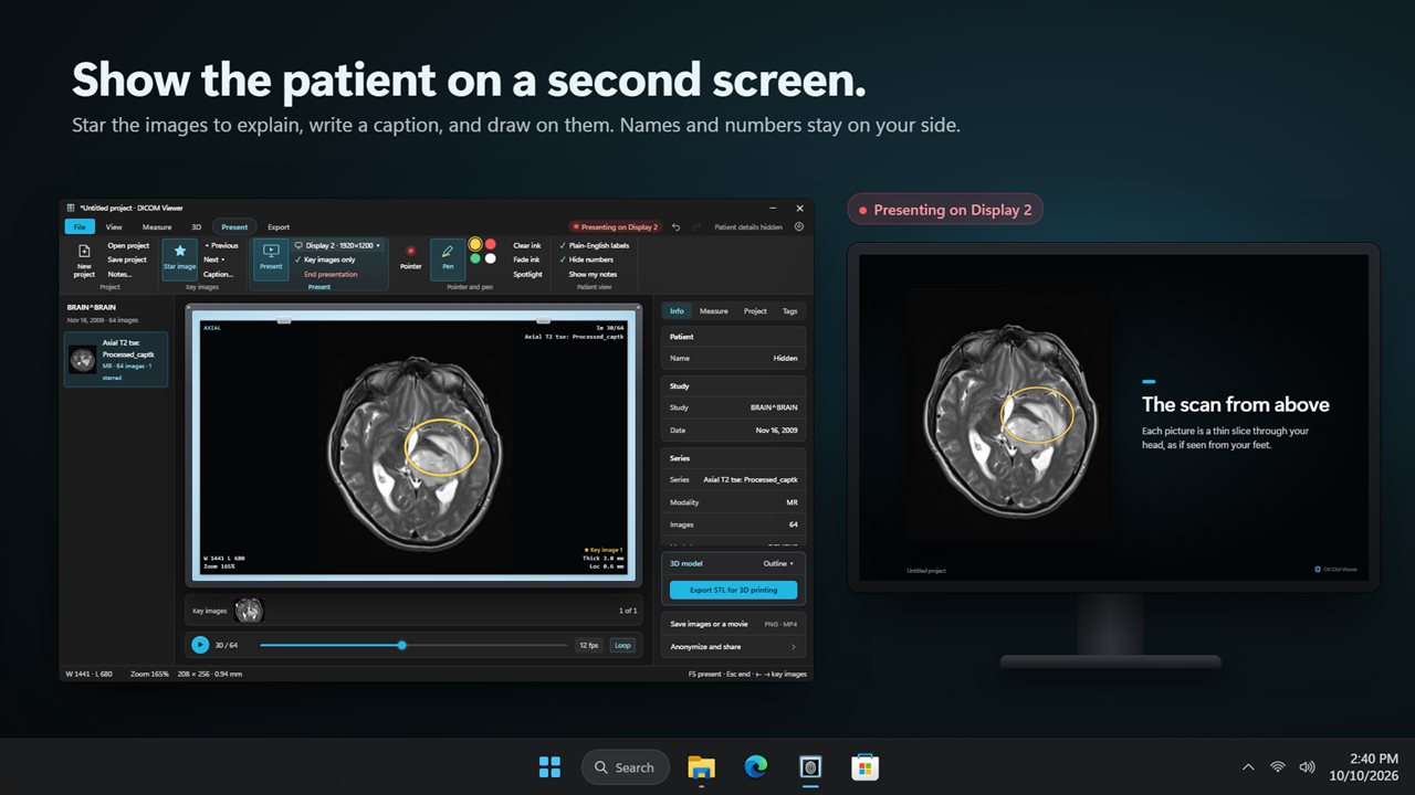
Walk a patient through their scan on a second screen.
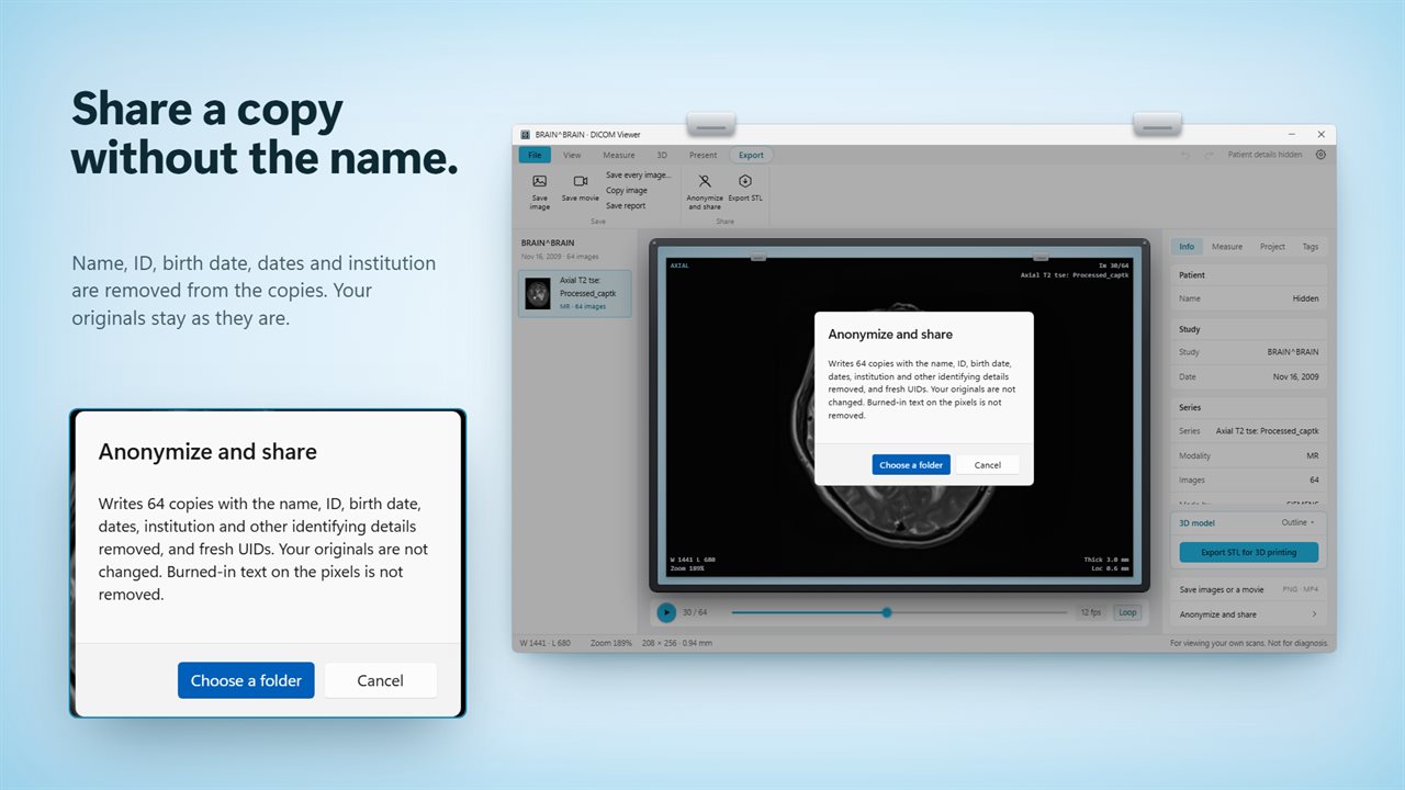
Share an anonymized copy without the patient's name.

CT, MRI and X-ray scans, every plane side by side with a 3D model.

190,000+downloads of EvlarSoft apps
Signed by EvlarSoftVerified US publisher
14-day money-backNo questions asked
Updates includedNo upgrade fees

A DICOM viewer for the scan disc you were handed

Hospitals and imaging centers hand out scans as DICOM files, usually on a CD with a DICOMDIR index, and Windows has nothing built in that can read them. A DICOM viewer fills that gap. This one is built for the person holding the disc as much as the specialist.

Choose Open disc and it follows the DICOMDIR; if a disc has no index, it reads each file on it instead, then groups the images into series in a side list. A copied folder, a loose image or a file missing its extension opens the same way, and dragging onto the window works as well. Files on the disc that are not DICOM images, such as the bundled viewer program, are skipped, and a note tells you how many.

Hide patient details is on from the start, so names, IDs and birth dates stay off the films, the info panel and any image you save until you turn it off. That makes it safer to look at a study with someone beside you or to share your screen.

Treat it as a way to look, never as a way to diagnose: the software has no regulatory clearance as a medical device, and nothing it shows should drive a clinical decision. If the images are yours, talk them through with your doctor.

How to open DICOM files and DCM scans on a PC

Whether the study came on a disc, a USB stick or as a download, the steps are short:

  • Insert the disc, then choose Open disc… and point it at the drive.
  • For a copied folder, choose Open folder…; for one or two images, Open files… accepts .dcm, .dicom, .dic and .ima.
  • Pick a series in the side list and turn the mouse wheel to move through the slices, or switch the wheel to zoom in Settings.
  • Press play to animate the stack at 5 to 30 frames a second, with Loop on or off.

An MRI and CT scan viewer that rebuilds every plane

Every CT or MR study arrives as a stack of flat slices. Turn on MPR + 3D and the viewer stacks them into a volume, then shows front and side views next to the original, with reference lines marking where each plane cuts the others. Thick-slab projection blends 2, 5, 10, 20 or 40 mm of slices as a maximum intensity (MIP), minimum intensity (MinIP) or average view.

For CT, one-click window presets cover lung, bone, soft tissue, brain, subdural and stroke, and the pixel value under the cursor is read out in Hounsfield units. You can also drag over an area to set the window from it, or type a custom width and level. Put 1, 2, 4 or 9 films on screen at once, scroll them together, or open a second study to compare against.

Turn a DICOM scan into an STL file for 3D printing

The same volume becomes a surface model you can drag to spin. CT models are cut at a Hounsfield threshold: bone at 300 HU, dense bone at 700 HU or skin at −300 HU, or any value you enter. MRI has no fixed scale, so the viewer offers an outline and an inner-detail level measured from the scan in front of you. Model detail runs from Draft to Fine.

Export the result as an STL model for a slicer or another 3D app. A series needs at least 8 evenly stacked slices with position data for this to work, so a single X-ray will not become a model.
Inside the viewer

  • Distance, angle and Cobb angle tools, plus rectangle, ellipse and freehand areas with mean, SD, minimum and maximum
  • Calibrate an image without pixel spacing by drawing along something of known length in millimeters
  • Arrows, notes and running spine labels drawn on the film
  • A measurement report saved as CSV or a text file, or copied to the clipboard
  • A tag panel listing the header of each image, with a filter box
  • Present mode with F5Starred key images and captions on a second screen, with pointer and pen
  • Projects that keep key images, captions, measurements and private notes for next time
  • Anonymize and shareCopies written with identifying details removed and fresh UIDs

Who uses it

Patients with a disc

See what was scanned before a follow-up appointment, and keep a PNG of the image your doctor pointed to.

Clinicians explaining a result

Star the key slices, write a caption a patient can read, and walk through them on a second monitor without names or IDs on show.

Makers and students

Build a bone or skin surface from a CT series and print it, or measure anatomy on teaching datasets.

Questions

Anything else? Ask us

Can it open the CD I got from the hospital?

Yes. Choose Open disc and it reads the disc's DICOMDIR index, or every image on the disc when there is none, and sorts the study into series. Folders, single files and DICOMDIR files open too, and you can drop them on the window.

Which compressed DICOM images does it open?

JPEG baseline and extended, JPEG lossless, JPEG-LS, JPEG 2000 and RLE, as well as uncompressed little endian, big endian and deflated files.

Why is 3D or MPR not available for my scan?

The front, side and 3D views need a CT or MRI series of at least 8 parallel slices of the same size, with position information. A single X-ray or a short or unevenly stacked series cannot be rebuilt.

Does Anonymize change my original files, and does it remove everything?

No to both. It writes copies to a folder you choose, with the name, ID, birth date, dates, institution and other identifying details removed and fresh UIDs, and leaves your originals alone. Text burned into the image pixels is not removed, so check the copies before sharing.

Can I use it to diagnose a scan?

No. It is not a medical device, it has not been cleared by any regulator, and its views, measurements and 3D models must not be used for any clinical decision. If you are a patient, ask your doctor about your images.

What can I save or export?

Images as PNG, a series as an MP4 movie, or WebM where MP4 is not available, the 3D model as an STL file, and measurements as CSV. It does not save to JPEG or write edited DICOM files, apart from anonymized copies.

What is a DCM file?

A DCM file is a single DICOM image: the picture plus a header with the patient, the study and how the image was taken. A full scan is usually hundreds of them, which is why a viewer groups them into series.

Can I convert a DICOM scan to a 3D model?

Yes, for CT and MRI series of at least 8 parallel slices. Turn on MPR + 3D, pick a surface level, and save the model as an STL file.

Can I look at two scans side by side?

Yes. Choose a layout of 2, 4 or 9 films for series in one study, or open a second study and compare it against the first.

Which versions of Windows does it run on?

Windows 10 (version 1809 or later) and Windows 11.

Is it a subscription?

No. DICOM Viewer is a one-time purchase of $29.99, and updates are included.

How do I get it?

Buy it on this page and your license key arrives by email. If the button says Notify me, DICOM Viewer is not on sale here yet; leave your email and we will tell you when it is.